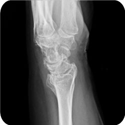
Western Health Orthopaedic Registrar Presentation – Distal Radius Fractures
Definition
- Fracture of the distal radius
Eponyms
- Colles (1814)
- extraartic fracture of distal radius with dorsal displacement of distal fragment
- Barton (1838)
- a fracture / dislocation or subluxation in which the rim of the distal radius, dorsally or volarly, is displaced with the hand & carpus
- Smith (1854)
- a volarly angulated Colles fracture
- Chauffeur
- fracture radial styloid
Incidence
- 1/6 of all fractures seen in emergency rooms
- ¾ of all forearm fractures
- Bimodal distribution: 6-10 & 60-70; in the second peak women substantially outnumber men
Aetiology
- FOOSH:
- Wrist at 40 – 90° dorsi-flexion usually produces fractures of the distal radius
- scaphoid fractures occur at about 97o
- When the wrist is in palmar flexion the stresses are reversed causing Smith type fractures or palmar peri-lunate dislocations
Anatomy
- There are three articular facets on the distal end of the radius:
- Scaphoid facet
- Lunate facet
- Sigmoid notch
- About 80% of the load from the carpus is borne on the scaphoid & lunate facets & about 20% on the triangular fibrocartilage
- With ↑ dorsal angulation of the distal radius more weight is borne on the TFC, which can result in pain at the radiocarpal articulation
Classification
- Older 1965
- Frykman 1967
- Fernandez 1987
- Melone
- Cooney
- Universal
- AO
Pathology
Function contrasted with radiological followup:
- Function is impaired if
- More than 20° of dorsal angulation
- Less than 11° of radial inclination
- More than 2mm of radial shift
- In young patients (under 25) loss of palmar tilt can be associated with mid carpal instability
Function & intra-articular involvement
- articular involvement in low energy fractures in older post menopausal women has little effect on the generally favourable outcome in these patients
- In younger patients it is important to reduce the intra-articular fracture to within 2mm or symptomatic post-traumatic arthritis will develop
History
Examination
Investigations
Xrays
- Three important radiographic measures
- Palmar slope of the distal end of the radius
- measured on the lateral
- averages 11 to 12°
- Radial inclination
- measured on the AP
- averages 22-23°
- Radial length
- measured on the AP, between the tip of the radial styloid process & the distal articular surface of the ulnar head
- averages 11-12 mm
- Palmar slope of the distal end of the radius
Treatment
Aim
- to restore radial length, inclination & tilt
- Acceptable angulation of the distal radius
- less than 2mm shortening
- 15o loss of normal volar angulation of the articular surface
- Accurate restoration of the articular surface is the most critical factor in achieving a successful result following intra-articular fractures of the distal radius
- residual incongruity
- more than 90% incidence of arthritis compared to only 11% where the joint was congruous
- residual incongruity
Stable fractures
- These are generally managed with closed reduction & immobilization in a plaster cast
- position of the forearm in immobilization, & extension of the cast past the elbow, doesn’t appear to influence the outcome of the wrist
- In theory the position of supination advocated by Sarmiento holds the DRUJ in a reduced position & minimizes the tendency of the brachioradialis to cause the distal fragment to displace in a radial direction
- Two studies have shown that there is lasting improvement in 33% & 54% of remanipulated fractures
Unstable fractures
- Percutaneous pinning
- Pins & plaster (incorporating the pins in the plaster)
- This has a very high complication rate & is no longer advocated; it was initially championed by Green
- External skeletal fixation
- This also has a high rate of complications including:
- Pin track infections
- Radial sensory neuritis
- RSD
- Wrist stiffness (particularly worries Damian Ryan)
- Fracture through pin sites
- It is significantly more effective in maintaining reduction for unstable fractures than is closed reduction & POP immobilization
- Radial length & inclination are usually re-established but palmar tilt is rarely restored to normal. This may be because the stout palmar radiocarpal ligaments are pulled out to maximal length before the dorsal radiocarpal ligaments which are Z-shaped
- This also has a high rate of complications including:
- Limited open reduction
- Technique intended for fractures where the radial styloid & scaphoid facet have been reduced closed by ligamentotaxis
- A dorsal incision is made & the lunate facet elevated, then supported with K wires plus or minus autogenous iliac crest bone graft, while an external fixation frame is applied. (Described by Axelrod)
- ORIF
- Advocated for two main groups:
- Two part shear fractures (Barton type)
- Complex intra-articular fractures which are not amenable to reduction by more limited approaches
- Advocated for two main groups:
Complications

In a large retrospective study of 565 fractures Cooney found a complication rate of 31%. Complications include:
1. Median nerve dysfunction
2. Malunion
3. Arthritis
4. Stiffness of the digits
5. EPL rupture
6. RSD
7. Volkmann’s ischaemic contracture
Median nerve dysfunction
1. If a complete nerve lesion doesn’t improve after reduction of the fracture the nerve should be explored
2. If there is a partial lesion of the nerve, the fracture should be reduced & the patient observed for 7 days, & the nerve explored if there is no change & some motor weakness
3. If a nerve lesion develops after reduction the splint should be removed & the wrist placed in a neutral position
4. If a nerve lesion exists in a patient who needs open intervention the nerve should be released at the same time
Tendon rupture
1. EPL is the tendon most often ruptured & this is due to ischaemia rather than attritional rupture on an osseous spike
2. In younger patients with high functional demands it is best treated with a tendon transfer from the adjacent extensor indicis
