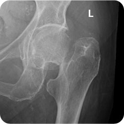
Definition
- fracture distal to articular surface & proximal to intertrochanteric region
Epidemiology
- 4ys younger than Intertrochanteric Fracture
- Averge age
- 77 yo F
- 72 yo M
Aetiology
Direct or Indirect
- 1 Direct blow Greater Trochanter
- 2 Post cortex impingement on rim
- 2° to ER
- Acts as a fulcrum
- 3 Bending Torque > Threshold
- Major trauma in young
- 4 Violent muscle contraction
- 5 Cyclical loading = Insufficiency fracture
Risks
- Osteoporosis
- Osteomalaica
- Co-morbidity
- Dementia
- Poor mobility
Classification
Undisplaced vs Displaced
- Undisplaced Garden I & II
- Displaced Garden III & IV
Pauwels
- Pauwels classification describes the angle the fracture makes with the horizontal.
- Garden felt that the fracture line was fairly constant at 50° & the obliquity of the fracture line was just a function of the XR plane.
| Type | Description |
|---|---|
| I | 30° from horizontal |
| II | 50° from horizontal |
| III | 70° from horizontal |
- Above is probably 2° to parallax errror
Garden
| Type | Description |
|---|---|
| I | Incomplete valgus impacted fracture |
| II | Complete undisplaced |
| III | Displaced with capsule intact Intact Weitbrecht Retinaculum Trabecular don’t line with acetabulum |
| IV | Displaced Trabecular line up with acetabulum |
- poor intra- & inter- observer reliability
- 22% of 100 femoral neck fractures were the fractures classified the same by 8 trained observers
- so the simpler classification of displaced vs. undisplaced is preferable
AO Classification of Subcapital Femoral Neck Fractures
| Group | Subgroup | Description |
|---|---|---|
| Type B1 subcapital fracture no or minimal displacement | B 1.1 | impacted in valgus > 15 degrees |
| B 1.2 | impacted in valgus < 15 degrees | |
| B 1.3 | nonimpacted | |
| Type B2 transcervical | B 2.1 | basicervical |
| B 2.2 | midcervical with adduction | |
| B 2.3 | midcervical with shear | |
| Type B3 displaced subcapital fractures | B 1.1 | moderately displaced in varus and external rotation |
| B 1.2 | moderately displaced with vertical translation and external rotation | |
| B 1.3 | markedly displaced |
Clinical Presentation
- Cause of fall
- TIA/ UTI / MI
- Pain
- Short & ER
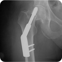
Management
- Based on age & displacement
- Treat comorbidity 1st
Undisplaced
- Internal Fixation
- 3 cannulated screws
- DHS ± Derotational Screw
- Nonunion Rate 5%
- AVN 10%
Displaced
- Controversial
- Options
- Reduction & Internal Fixation
- Nonunion 33%
- AVN 16%
- Conversion to salvage THR 30%
- results of salvage arthroplasty were markedly inferior to those of a primary arthroplasty for a displaced subcapital fracture.
- higher rates
- dislocation
- superficial infections
- revision THR
- overall prosthetic survival rate significantly lower.
- functional results were inferior.
- reason for poor results
- disuse osteoporosis
- muscle wasting
- extrusion of cement through screw holes.
- Hemiarthroplasty
- Principles
- Hemiarthroplasty leads to suboptimal outcomes in active older patients, with acetabular erosion & pain in 20%.
- Contraindications
- Medical conditions that affect potential fracture union & contribute to acetabular pathology
- Paget’s disease
- renal osteodystrophy,
- osteoporosis
- THR instead
- Medical conditions that affect potential fracture union & contribute to acetabular pathology
- Types
- Uncemented Unipolar Unimodular
- eg. Moore’s Hemiarthroplasty
- high revision rate for loosening
- eg. Moore’s Hemiarthroplasty
- Modular Hemiarthroplasty
- Unipolar
- Bipolar
- RCT
- no difference between the two
- pain
- satisfaction
- complication rate
- functional ability.
- no difference between the two
- Uncemented Unipolar Unimodular
- Principles
- Total Hip Replacement
- 90% of patients treated with THR report no or little pain after THR.
- Infection rates have been higher among patients undergoing arthroplasty using the posterior approach compared with other approaches to the hip in these patients.
- Dislocation rates are higher in patients who have acute THR.
- metaanalysis
- dislocation rates
- 2.1% for unipolar hemiarthroplasty
- 2.9% for bipolar hemiarthroplasty
- 10.7% for total hip arthroplasty
- dislocation rates
- Reoperation rates
- lowest for all treatment options (12% at 3 to 6 years).
- Reduction & Internal Fixation
Operative Technique
Closed Reduction & Internal Fixation
- Anatomical Reduction
- Optimum reduction of the femoral neck fracture has been shown, in numerous studies, to be associated with a lower rate of avascular necrosis of the femoral head.
- If AVN does develop, there is no evidence that core decompression is beneficial
- If these complications are avoided, these patients do better than arthroplasty patients of any kind.
- Urgent procedure
- Urgent reduction of femoral neck fractures has been shown to reduce the risk of avascular necrosis in patients undergoing internal fixation of displaced femoral neck fractures.
- However, mortality & morbidity rates have been shown to be higher when the medical condition of these patients is not optimised before surgery.
- Compressive screws
- Traction Table & II
- Reduction Techniques
- Type 1
- flex the hip to 45° while it is in slight abduction
- extend the hip while gently increasing traction
- then internally rotate it to 30 to 45° in full extension
- It is recommended that if this manouver, which should rely primarily on extension & internal rotation, does not reduce the hip satisfactorily under fluoroscopic control, then the surgeon should proceed to an open reduction rather than engage in repeated attempts with greater force, which could damage the blood supply to the femoral head.
- Leadbetter Manoeuvre
- Flex/Traction/ IR /Ext /ABD
- the hip is fully FLEXED
- and slightly ADDUCTED as TRACTION is applied.
- Full INTERNAL ROTATION is then applied.
- leg is then CIRCUMDUCTED into slight abduction while the rotation is maintained
- then brought into EXTENSION
- Some authorities believe that while the XR may look reduced the head has actually rotated on the neck, & there is a higher risk of vascular problems.
- Flex/Traction/ IR /Ext /ABD
- Type 1
- Accept only slight valgus
- After reduction, the outline of the head on the neck should present a gentle S on every view.
- Any varus predisposes to nonunion; excessive valgus leads to ↑ AVN.
- Anteversion or retroversion of more than 20° has been shown to ↑ the rate of nonunion.
- Posterior comminution of the femoral neck ↑ nonunion.
- Note on positioning: extension & internal rotation (the position for fixation) ↑ pressure within the joint capsule.
- Consider capsulotomy to ↓ pressure
- No proven benefit
- Internal Fixation
- 3 cannulated screws, in parallel.
- More than 3 screws is not biomechanically advantageous
- ↑ the risk of pin penetration.
- 3 cannulated screws, in parallel.
Open Reduction
- Take off Traction Table
- Lateral decubitus position on normal table
- Watson Jones approach
- straight lateral incision
- interval
- tensor fasciae lata & gluteus medius muscle
- bluntly down to the anterior aspect of the hip capsule
- origin of the vastus lateralis is elevated from the intertrochanteric ridge
- elevation of the vastus lateralis
- incision of the capsule
- split capsule over the femoral neck anteriorly
- dissect off the intertrochanteric ridge one centimetre inferiorly & one centimetre superiorly
- insert a small, pointed Hohmann retractor extracapsularly onto the anterior part of the acetabular rim to expose fracture
- reduction
- bone-hook is placed onto the greater trochanter
- unscrubbed assistant who is outside of the sterile field brings the hip into external rotation.
- With lateral traction by means of the bone-hook, the fracture is disimpacted.
- femoral head, which nearly always lies posteriorly, is then lifted anteriorly (the shaft is actually translated posteriorly) by a dull, curved instrument, as the lateral traction is released & the limb is placed back into maximum internal rotation.
- reduction is held provisionally by a 2.0-millimeter Kirschner wire.
- reduction should then be confirmed visually, by palpation, & on both anteroposterior & lateral radiographs.
Complications
 Cut Out of DHS
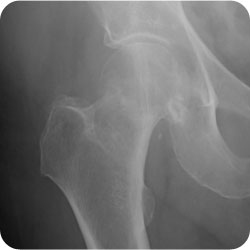 Type 1 Subcapital NOF
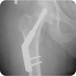
- 14-36% for 1y >65yo
- 5%
- 12% if displaced
- Undisplaced <10%
- Displaced = 20-35%
- Most commmonly Anterior Superior Lateral
Risks
- Delay in Reduction
- Poor Reduction
- Displacement
- Injury velocity
Revascularization
- Relies on
- Existing Medial Epihyseal Blood Vessels
- Remaining Lateral Epiphyseal Blood Vessels
- Metaphyseal Blood Vessels crossing fracture
- Decreased by
- Non- Union at fracture
- Mal-reduction
- Last area to regain supply is superolateral subchondral region
- 1st sign = Joint collapse
- Minimal ~ 6/12
- Most ~ 1yr
- Max ~ 5 yr
- 1st sign = Joint collapse
- If patient immobilised, may see relative ↑ in density of dead bone
- Less common with ORIF & early mobilisation
- Role of prognostic MRI/ TE Scan uncertain
- Prognosis value limited
- ? Scan at 1/12
- Only 30% with AVN will need reoperation