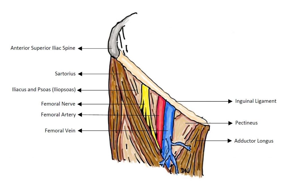
Iliac fossa, entering thigh beneath the lateral part of inguinal ligament
Insertion
Inserted in front of psoas tendon & small area of femoral shaft, just below the lesser trochanter
Nerve Supply
Femoral nerve (L2,3) in iliac fossa
Action
Powerful flexor of hip
Psoas major
Origin
Lumbar spine, passes deep to middle of inguinal ligament
Insertion
Lesser trochanter
Both iliacus & psoas pass across the front of the capsule of the hip joint, with the bursa intervening
burse may communicate with the joint thorugh a gap in the capsule that lies between the iliofemoral & pubofemoral ligaments
Nerve Supply
First 3 lumbar nerves (mainly L2)
Action
Powerful flexor of hip
Pectineus
Quadrilateral muscle
Covered anteriorly by infolding of fascia lata
Femoral vein & canal lie on top of it
Adductor brevis & anterior division of obturator nerve lie behind it
Origin
Pectineal line of pubis & narrow area if bone below
Insertion
Vertical line below lesser trochanter
Nerve Supply
Anterior division of femoral nerve (L2,3)
Occasionally twig from obturator nerve (L2,3)
Action
Flexes & adducts thigh
Quadriceps femoris
Nerve Supply
each muscle is supplied by its own branch from the femoral nerve (L3, 4)
Action
main extensor of knee
rectus femoris can assist iliopsoas flex the hip
Rectus femoris
Origin
Arises ilium by 2 heads
Reflected head – groove above acetabulum
Straight head – upper half of AIIS, above iliofemoral ligament
2 heads unite to form the anterior lamina of quadriceps tendon
Vastus Lateralis
Origin
Extensive linear origin from upper part of intertrochanteric line, greater trochanter, lateral lip of linea aspera of femur, lateral intermuscular septum
descending branch of lateral circumflex artery & nerve to vastus lateralis lie between vast lateralis & intermedius
Vastus intermedius
Origin
ant & lateral surfaces of upper 2/3 of shaft of femur
Articularis genu
Origin
ant surface if lower femoral shaft, deep to vastus intermedius
Insertion
upper convexity of suprapatellar bursa
Vastus medialis
Origin
Lower part of intertrochanteric line, medial lip of linea aspera, tendon of adductor magnus below the hiatus for femoral vessels
Action
Lowest fibres are indispensable for stability of patella