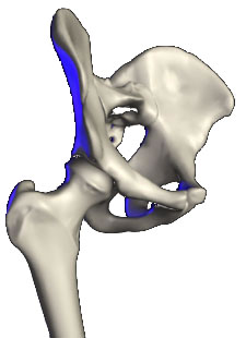
Articulation of the Hip Joint

- multiaxial ball & socket synovial joint
- Acetabulum
- formed by the fusion of three bones, the ilium, ischium & pubis
- These meet at a Y-shaped cartilage which forms their epiphyseal junction. (closes after puberty)
- acetabular articular surface is covered by hyaline cartilage, & is a C-shaped concavity
- It’s peripheral edge is deepened by the fibrocartilagenous acetabular labrum which further encloses the femoral head, increasing stability
- labrum is continued across the acetabular notch as the transverse acetabular ligament, which unlike the labrum, does not have any cartilage cells
- transverse ligament gives attachment to the head of the femur, via the ligamentum of teres
- central nonarticular area of the acetabulum is occupied by the haversian fat pad, which is covered by synovial membrane
- Head of femur
- spherical head of the femur forms 2/3 of a sphere
- completely covered by hyaline cartilage, accept at it’s fovea, with a little encroaching on the anterior neck for articulation in flexion
- More than half of the head is contained by the acetabulum
- pit or fovea, is for attachment of the ligament of the head of the femur, which arises from the margins of the acetabular notch, & prevents excessive external rotation
- neck of the femur is narrower than the head, allowing considerable movement in all directions before the neck impinges
- This feature is important in giving the hip a wide range of movement
- femoral neck forms approximately 125º angle of inclination with the shaft. (greater in children)
- 12-15º anteverted. (40º at birth)
- Capsule
- attached around the labrum & transverse ligament, then passes laterally like a sleeve to attach to the neck of femur
- In front it attaches to the intertrochanteric line, but posteriorly it extends only halfway along the neck
- From these attachments the fibres of the capsule are reflected back along the neck of femur, blended with the periosteum, to the articular margin of the head
- These are the retinacular fibres which bind the nutrient vessels, chiefly from the trochanteric anastomosis, to supply the head.
Ligaments
- The capsule is strengthened by three ligaments which arise from each bone of the hip, which “unwind” with the hip flexed & externally rotated (fetal position) to lie parallel with the neck & relaxed. Thus extension & medial rotation tightens these
- iliofemoral ligament (Bigelow)
- is the strongest, & Y-shaped
- stem of the Y arises from the lower half of the anterior inferior iliac spine & the rim of the acetabular rim
- The diverging limbs are attached to the upper & lower of the intertrochanteric line.
- This limits extension, & acts as a fulcrum around which the neck rotates when the head dislocates
- In standing, as the pelvis is thrust anteriorly, much of the body weight is borne by these ligaments
- pubofemoral ligament
- passes from the iliopubic eminence & obturator crest to the capsule on the inferior part of the neck of femur, combines with the iliofemoral ligament
- Thus it prevents over abduction of the hip
- ischiofemoral ligament
- weakest
- arises from the posterioinferior margin of the acetabulum, passing laterally to the capsule, spiraling upwards to a band of fibres that run in the capsule transversely around the neck of femur
- forms the zona orbicularis , which gives the capsule an hourglass appearance
SYNOVIAL MEMBRANE
- attached to the articular margins around the labrum & transverse ligament
- lines the capsule, being reflected back along the neck, where it invests the retinacular fibres
- haversian fat pad & ligament of the head are also invested by a sleeve attached to the margins of the fovea & concavity of the acetabulum
- In 10% a perforation in the anterior capsule between iliofemoral & pubofemoral ligaments permits communication between the synovial cavity & iliac bursa
- Anteriorly
- lies the iliac bursa, which lies beneath the iliacus, extending to the iliac fossa. The psoas tendon separates the capsule from the femoral artery, & the iliacus from the nerve, while medially the pectineus lies between the capsule & femoral vein
- Superiorly
- overhangs the gluteus medius, & inferiorly the obturator externus spirals around the neck
- Posteriorly
- lies the piriformus, the obturator internus & gemelli
NEUROVASCULAR
- The head & intracapsular neck receive their supply from the trochanteric anastomose, mainly through branches of the medial circumflex vessel
- The artery in the ligament of the head comes from the posterior branch of the obturator artery, & usually atrophies by the age of 7
- The three nerves of the pelvic girdle supply the joint, the femoral via the nerve to rectus femoris, the sciatic via the nerve to quadratus femoris, & the obturator from it’s anterior division
MOVEMENTS
- The articulation of the femoral head in the acetabulum permits 3° of rotational freedom about the pelvis
- There is essentially no translation in the hip
- Flexion
- head of the femur rotates about a transverse axis that passes through both acetabular, normally about 135º, limited by the thigh on the abdomen
- muscles are psoas major & iliacus, assisted by rectus femoris, tensor fascia latae, sartorius & pectineus
- Extension
- gluteus maximus at the extremes of movement
- hamstrings in the intermediate stage
- limited by tension in the iliofemoral ligament at about 30º
- Abduction & adduction
- With this movement the femoral head rotates in the AP plane.
- Adduction
- 25 is produced by the pectineus, adductors longus, brevis, magnus & gracilis
- This is limited by tension in the glutei
- Abduction
- gluteus medius, & minimus, & assisted by the piriformus
- limited to 45º by the tension in the pubofemoral ligament
STABILITY OF THE HIP
- Bony factors & the iliofemoral ligament are mainly responsible, with the gluteus medius & minimus responsible with the hip in motion
- capsule of the hip is one of the strongest in the body
KINETICS
- The range of excursion & power of the muscles about the hip are ↑ by the length of the neck, prominence of the trochanters, as well as the long moment arms from their positions relative to the centre of the joint
JOINT FORCES
- during the single leg stance, the forces transmitted across the hip joint are estimated to be between 2 – 2.8 times the body weight
- During two legged stance, forces have bee estimated to be half this
- During gait these forces can be as high as 3 times body weight, mainly during the foot strike, with the forces half this during the swing phase
