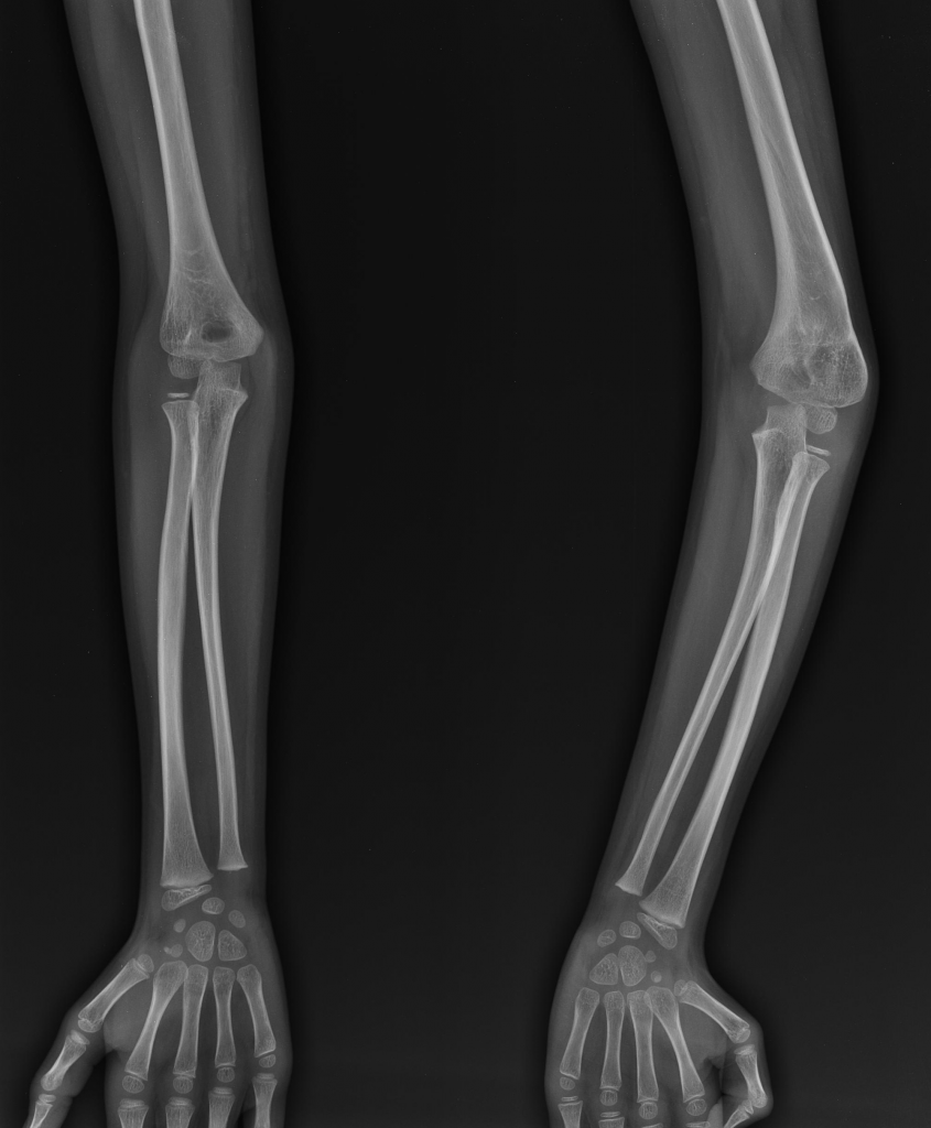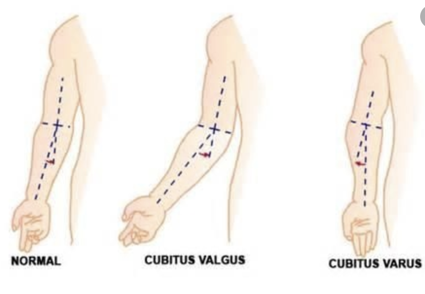

Definition:
In the erect anatomic position, the carrying angle of the elbow is angulated medially (towards the body), in the coronal plane. Also commonly referred to as “gunstock deformity.
Anatomy:
Normal anatomical carrying angle of humerus 5-15 degrees
Trochlea tilted approximately 4-degrees valgus in males, 8 degrees in females.
Trochlea 3-8 degrees externally rotated.
Epidemiology:
Wide ranging data on the incidence of cubitus varus in paediatric papers.
Published incidence rates from 4%-58%.Commonly quoted at 10%.
Aetiology:
Cubitus varus has historically been associated with injury to the distal humerus epiphysis and subsequent growth disturbance. This is still felt to be a cause.
More recent thinking is regarding mal-reduction and malunion, with extension, internal rotation and medial displacement of the distal aspect of the fracture.
Pathology:
- While supracondylar fractures are the most common cause for cubitus varus, other causes are reported
- Medial epiphyseal injury or infective process
- Osteonecrosis of the trochlea
- Epiphyseal dysplasia (congenital)
- Neoplasia
Natural History:
Deformity does not correct with time and remodelling.
Can lead to long term posterolateral rotatory instability.
History:
Previous elbow fracture – commonly supracondylar
Functional deficit in rotation can be noted – using computer mouse
Examination:
Bony prominence – lateral condyle
Varus carrying angle of the elbow
Flexion limitation
Wasting of musculature
Important to undertake careful neurovascular exam – reported cases of tardy ulna nerve palsy (less common than in cubitus valgus)
Investigations:
X-ray – assessing Baumann’s angle. Normal range is 64-81 degrees. Increased in cubitus varus.
CT with 3D reconstruction may aid operative planning
Treatment:
- Non-operative management if no functional deficit and acceptable cosmetically
- Lateral closing wedge osteotomy of the distal humerus
- Medial opening wedge osteotomy of the distal humerus
- Oblique osteotomy with de-rotation
- Complex osteotomies such as dual plating for extreme deformity
- Dome osteotomy
- Guided growth (age dependant)
References
Photo reference – https://www.quora.com/Is-Cubitus-varus-a-permanent-rejection-in-the-armed-forces-for-NDA-entry
