Definition
- Fracture through the distal humerus just proximally to the growth plate
Aetiology
- Most commonly fall on outstretched hand with extended elbow-
- Extension injury with posterior fragment displacement most common ~98% of all injuries
- Rarely flexion injuries can occur with anterior fragment displacement ~2% of all injuries
Epidemiology
- Most common fracture of the elbow in kids accounting for approximately (3%) of all paediatric fractures
- Peak incidence age 5-7 years old
Anatomy
| Ossification Centre | Ossification (Years Old) | Fusion (Years Old) |
| C- Capitellum | 0 – 1 | 10 – 15 |
| R- Radial head | 2 – 6 | 12 – 16 |
| I- Internal (Medial) Epicondyle | 2 – 8 | 13 – 17 |
| T- Trochlea | 5 – 11 | 10 – 18 |
| O- Olecranon | 6 – 11 | 13 – 16 |
| E- External (Lateral) Epicondyle | 8 – 13 | 12 – 16 |
On average girls will develop ossification and fusion of these sites prior to boys by about 1-2 years.
Pathology
- 95 % are hyperextension type with olecranon as the fulcrum
- rotation leads to instability & tilting
Classification
Gartland Classification
- Lateral radiograph is used to determine the staging for this classification.
| Type | Description |
|---|---|
| 1A | Non or minimal displaced |
| 1B | Non displaced with medial impaction |
| 2A | Displaced with angulation however posterior cortex intact. |
| 2B | Displaced with angulation however posterior cortex intact. Some degree of rotatation or translation of fracture present. |
| 3 | Complete displacement ( usually posteromedial) |
- Further classification should include other features (extension/flexion) and any neurological/vascular involvement
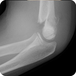 Type II Supracondylar Fracture
History
- Pain and swelling of affected elbow
- Decreased use/function
- Fall on to outstretched hand with straight elbow
Examination
- Presents with painful swollen elbow
- Neurological assessment
- AIN most commonly affected nerve in extension injury (AIN>median>radial>ulnar)
- Ulna most commonly injured in flexion injury
- Vascular status
- Distal Pulses
- Peripheral perfusion (eg CRT)
- Often categorised as normal, pulseless with pink hand (pulseless but CRT retained) or pulseless with white hand (dysvascular)
- Emergency surgery is indicated for pink pulseless hand and dysvascular presentations
- Examination can be difficult due to age of patient and pain/fear. Comprehensive examination and documentation of neurovascular status should however always be performed.
Investigations
- X-Ray
- best investigation to assess for fracture
- AP/Lateral images
- if no fracture visible, the presence of a posterior fat pad on XR can indicate the presence of an underlying fracture
- anterior humeral line
- measured from lateral humeral XR
- anterior humeral line is a marker drawn down the anterior humerus, should intersect with the middle third of the capitellum ossification site (may be in the anterior third in kids under 4)
Differential Diagnosis
- Lateral condyle fracture
- second most common paediatric fracture (12-20% of all paediatric fractures)
- highly associated with missed diagnosis causing long term issues with non union/mal union
Treatment
- Type 1A
- above elbow cast for 3 weeks
- Type 1B
- above elbow cast for 3 weeks
- if significant varus exists manipulate & if manipulated pin it
- Type 2A
- Debate exists to whether casting or percutaneous pinning is most appropriate
- Percutaneous pinning growing favour of treatment however casting may still be viable
- Type 2B
- Closed reduction and percutaneous pinning
- Type 3
- Closed reduction & percutaneous pinning is the standard
- Open reduction is rarely required
Surgical Technique
- Considerations
- Capitellum should not be posterior to the anterior line of the humerus
- Closed reduction & percutaneous pinning is the standard
- Open reduction is rarely required
- Medial versus lateral incision
- S shaped anterior incision if vascular repair is required
- Medial displaced – pronate
- Laterally displaced – supinate
- Technique
- GA
- Traction to disengage the proximal fragment from the brachioradialis
- Then fix the translation in the coronal plane
- Correct rotational deformity
- Follow this with bringing the olecranon back over, like reducing an elbow dislocation
- Bring the arm out in extension & pronate or supinate
- Now pin lateral & then extend elbow for medial – 0.062 pins
- Assess an adequate Reduction:
- Anterior humeral line bicects the capitellum
- Bowman angle within 5° of the contralateral side
- diameter of the fracture ends are equal
- If pulseless – reduce, then reassess pulse if viable pin the arm, if not viable pin the arm & follow with vascular repair
- Late cubitus varus is treated with an osteotomy
Complications
- Chance of ipsilateral distal radius fracture
- supracondylar fractures with associated diaphyseal fractures are at an increased risk of compartment syndrome
- Neurovascular compromise
- Traumatic
- AIN>median>radial>ulnar
- Iatrogenic
- Often ulna nerve injury
- Most recover within 2-3 months
- Traumatic
- Cubitus Varus
- most common complication as result of fracture
- potential complication regardless of casting or pinning
- painless complication however may be linked with increased risk of elbow instability later in life
- for children with substantial residual cubitus varus, lateral osteotomy should be considered
- Pin Site Infections
- ~1-2% of pinning operations
Prognosis
- With correct diagnosis and appropriate treatment good outcomes are expected with minimal long term issues or complications
- Delays in identifying and treating neurovascular compromise however can cause long term issues for the patient, therefore thorough examination and early involvement of orthopaedics is required
T Condylar Fracture
- Adolescent type fracture & treat with fixing the articular surface first
References
- Abzug, Joshua M. MD; Herman, Martin J. MD Management of Supracondylar Humerus Fractures in Children: Current Concepts, Journal of the American Academy of Orthopaedic Surgeons: February 2012 – Volume 20 – Issue 2 – p 69-77 doi: 10.5435/JAAOS-20-02-069
- American Academy of Orthopaedic Surgeons EvidenceBased Clinical Practice Guideline for the Treatment of Pediatric Supracondylar Humerus Fractures
- Kumar V, Singh A. Fracture Supracondylar Humerus: A Review. J Clin Diagn Res. 2016;10(12):RE01-RE06. doi:10.7860/JCDR/2016/21647.8942
- Author Contribution
- Miyazaki CS, Maranho DA, Agnollitto PM, Nogueira-Barbosa MH. STUDY OF SECONDARY OSSIFICATION CENTERS OF THE ELBOW IN THE BRAZILIAN POPULATION. Acta Ortop Bras. 2017;25(6):279-282. doi:10.1590/1413-785220172506170954
- Tejwani, Nirmal MD; Phillips, Donna MD; Goldstein, Rachel Y. MD, MPH Management of Lateral Humeral Condylar Fracture in Children, American Academy of Orthopaedic Surgeon: June 2011 – Volume 19 – Issue 6 – p 350-358
Author Contribution
Updated by Dylan Ellis 2021
Reviewed by